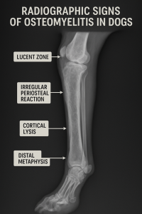
Radiographic Signs of Osteomyelitis in Dogs
- Periosteal Reaction (Osteogenesis, New Bone Formation):
- New bone formation along the outer surface of the affected bone.
- Manifests as irregular, fluffy, or laminated periosteal proliferation.
- Bone Destruction (Osteolysis):
- Evidence of bone destruction seen as areas of radiolucency or bone resorption.
- Can lead to cortical thinning or complete destruction of bone architecture.
- Soft Tissue Swelling:
- Visible adjacent to the affected bone.
- Indicates inflammation and edema in the surrounding tissues.


- Involucrum Formation:
- New layer of periosteal bone formed around necrotic bone in response to infection.
- Appears as a thin, irregular layer of new bone surrounding the affected area.
- Sequestrum Formation:
- Presence of a fragment of necrotic bone separated from healthy bone.
- Appears as a radiopaque focus within an area of bone destruction.
- Joint Involvement:
- If infection extends into the joint:
- Radiographic signs may include joint effusion.
- Periarticular osteopenia (reduced bone density around the joint).
- Irregular joint margins.
- If infection extends into the joint:
- Regional Osteopenia (osteoporosis):
- Decreased bone radiopacity (radiographic density) observed in the region affected by osteomyelitis,
Considering these radiographic signs alongside clinical symptoms (such as lameness, pain, and fever) and laboratory findings for an accurate diagnosis. Veterinary consultation is essential for proper diagnosis and treatment planning.
Alireza Ghadiri
Professor, DVM, DVSc
Board Certified Veterinary Radiologist
Smart TRCVET